 By
By 
When to Escalate Care for Low SpO2 Despite Supplemental Oxygen?
For over two decades in respiratory health, I've witnessed the profound anxiety that grips individuals and caregivers when a patient's oxygen saturation (SpO2) remains stubbornly low, even with supplemental oxygen. It's a critical moment, often fraught with uncertainty, where the right decision can mean the difference between stable recovery and rapid deterioration. My experience, spanning countless emergency room shifts, ICU rounds, and home health consultations, has taught me that while numbers are important, the complete clinical picture is paramount.
This persistent hypoxemia, where the body isn't getting enough oxygen despite intervention, presents a significant dilemma. It's not just a minor concern; it's a red flag indicating that the current treatment isn't sufficient or that the underlying condition is worsening. The challenge lies in accurately assessing the situation and knowing precisely when to transition from routine monitoring to urgent medical escalation, a decision that many find daunting.
In this definitive guide, I will share my expert framework and actionable insights to empower you with the knowledge needed to make informed decisions. We'll delve into the critical signs beyond just the SpO2 number, explore a practical escalation checklist, and examine real-world scenarios. My goal is to equip you with the confidence to understand when to escalate care for low SpO2 despite supplemental oxygen, ensuring timely and appropriate intervention.
Understanding Hypoxemia and Supplemental Oxygen
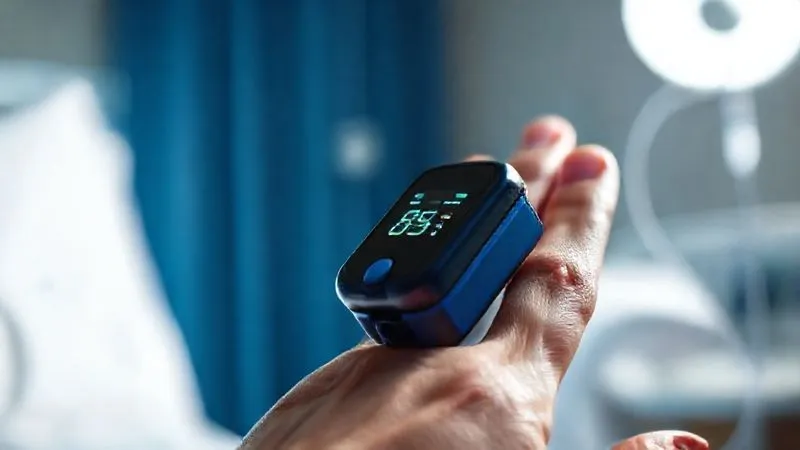
Oxygen saturation, or SpO2, measured non-invasively by a pulse oximeter, reflects the percentage of hemoglobin carrying oxygen in your blood. A normal SpO2 for most healthy individuals ranges between 95-100%. For those with chronic respiratory conditions like COPD, a target range of 88-92% might be considered acceptable, as their bodies have adapted to slightly lower oxygen levels.
Supplemental oxygen is a vital therapy designed to increase the amount of oxygen available for the body's tissues. It's prescribed for various conditions causing hypoxemia, from acute infections like pneumonia to chronic lung diseases. The expectation is that by providing extra oxygen, the SpO2 will rise to a safer, more functional level, alleviating symptoms of oxygen deprivation.
However, the core problem we're addressing today arises when this expected improvement doesn't occur, or worse, when the SpO2 continues to decline despite what appears to be adequate oxygen delivery. This scenario signals a critical failure in the body's ability to oxygenate, or a significant worsening of the underlying pathology, necessitating a rapid reassessment and often, an escalation of care. Ignoring these signs can have severe, even life-threatening, consequences.
The Critical Signs: Beyond the Numbers
While a low SpO2 reading is the initial trigger, an experienced clinician understands that it's merely one piece of a much larger puzzle. The true indicator of a patient's clinical status is the comprehensive assessment of their respiratory effort, mental state, and other physiological responses. Relying solely on the pulse oximeter can be misleading; it's the pattern and associated symptoms that truly tell the story.
Worsening Respiratory Effort
This is arguably the most crucial clinical sign. If a patient is working harder to breathe despite receiving supplemental oxygen, it's a clear signal of distress. Observe for:
- Increased Respiratory Rate (Tachypnea): Breathing faster than normal, especially if it's a sustained increase from their baseline.
- Accessory Muscle Use: The patient using muscles in their neck, shoulders, and abdomen to breathe, rather than just the diaphragm. Look for shoulders rising with each breath or the sternocleidomastoid muscles bulging.
- Retractions: The skin sucking in around the ribs (intercostal), above the collarbone (supraclavicular), or below the sternum (substernal) during inhalation. This indicates significant negative pressure needed to draw air in.
- Nasal Flaring: Widening of the nostrils with each breath, a common sign of respiratory distress, especially in children but also present in adults.
- Pursed-Lip Breathing: While often a compensatory mechanism in COPD, new or exaggerated pursed-lip breathing can indicate increasing effort.
Any of these signs, particularly when worsening over a short period, indicates that the body is struggling to maintain adequate oxygenation and ventilation, even with external support.
Altered Mental Status
The brain is highly sensitive to oxygen deprivation. When hypoxemia becomes significant, it can manifest as changes in a patient's cognitive function. These changes can be subtle initially but can progress rapidly.
- Confusion or Disorientation: Difficulty remembering things, not knowing where they are, or who people are.
- Lethargy or Drowsiness: Unusual sleepiness or difficulty staying awake and alert.
- Agitation or Restlessness: Paradoxically, some patients become agitated, irritable, or restless due to hypoxia, as the brain struggles to function normally.
- Slurred Speech: Difficulty articulating words clearly.
In my experience, a change in mental status, especially new-onset confusion or unusual drowsiness in a patient who was previously alert, is one of the most concerning signs of worsening hypoxemia. It often precedes other, more overt signs of respiratory failure and should always prompt immediate investigation.
Cyanosis and Skin Changes
Cyanosis, a bluish discoloration of the skin or mucous membranes, is a classic sign of severe hypoxemia, indicating a significant amount of deoxygenated hemoglobin. While it can be harder to detect in individuals with darker skin tones, careful observation is crucial.
- Central Cyanosis: Bluish discoloration of the lips, tongue, or mucous membranes inside the mouth. This is a sign of widespread hypoxemia.
- Peripheral Cyanosis: Bluish tinge to the fingertips, nail beds, or toes. This can be due to poor circulation (e.g., cold hands) but in the context of low SpO2, it can also indicate systemic hypoxemia.
- Pallor: Unusual paleness of the skin, often accompanied by clamminess, can indicate poor perfusion or shock, which can be linked to severe respiratory compromise.
Always assess these changes under good lighting and compare them to the patient's baseline if possible. The presence of new or worsening cyanosis, particularly central cyanosis, is a critical emergency.
Cardiovascular Compromise
The heart works harder when the body is deprived of oxygen. Initial responses might be compensatory, but sustained hypoxemia can lead to cardiac distress.
- Tachycardia: An abnormally fast heart rate (e.g., >100 beats per minute) is often an early compensatory response to low oxygen.
- Bradycardia: An abnormally slow heart rate (e.g., <60 beats per minute) can be a late and ominous sign of severe, prolonged hypoxemia, indicating cardiac exhaustion.
- Hypotension: A drop in blood pressure, potentially indicating shock, can occur with profound hypoxemia and respiratory failure.
- Arrhythmias: Irregular heart rhythms can be triggered by oxygen deprivation to the heart muscle.
Monitoring vital signs beyond just SpO2 is critical. A sustained or worsening trend in heart rate or blood pressure, especially when accompanied by low SpO2, demands urgent attention.
Persistent or Declining SpO2 Trend
It's not just about a single low reading; it's about the trend. A transient dip might not be as concerning as a sustained drop or a continuous decline, even if the absolute number isn't critically low yet. For example, a patient whose SpO2 was consistently 92% on 2 liters of oxygen, suddenly dropping to 88% and staying there despite increasing oxygen, signals a problem. Similarly, a patient who initially responded well to oxygen but then starts to trend downwards is deteriorating.
Understanding the patient's baseline and how they typically respond to oxygen is key. According to a comprehensive review in the New England Journal of Medicine, continuous pulse oximetry monitoring, with attention to trends, significantly improves early detection of respiratory compromise compared to intermittent checks. This emphasizes the importance of consistent observation rather than isolated data points.
| SpO2 Range | Clinical Action |
|---|---|
| 95-100% | Normal, continue routine monitoring |
| 90-94% | Mild hypoxemia, assess patient, ensure oxygen delivery is optimal |
| 85-89% | Moderate hypoxemia, increase oxygen, reassess quickly, notify medical team |
| <85% | Severe hypoxemia, immediate medical emergency, escalate care urgently |
My Framework for Escalation: The "ABCDE" Checklist
When faced with a patient whose SpO2 is low despite supplemental oxygen, a structured approach is essential to avoid missing critical steps. I've developed a simple yet effective framework over the years, the "ABCDE" Checklist, to guide immediate actions and decision-making.
- Assess Airway & Breathing: This is always the first step. Is the airway clear? Listen for stridor, gurgling, or wheezing. Observe the chest rise and fall. Is the patient breathing effectively? Check their respiratory rate and effort. A compromised airway or ineffective breathing will negate any amount of supplemental oxygen.
- Boost Oxygen Delivery: Ensure the oxygen delivery system is functioning optimally. Is the oxygen tank full? Is the flow rate set correctly? Is the nasal cannula or mask properly fitted and sealed, without kinks in the tubing? Consider increasing the oxygen flow rate or switching to a higher-delivery device, such as a non-rebreather mask, if appropriate and available. This is a crucial immediate intervention to see if the SpO2 responds.
- Consider Underlying Cause: While boosting oxygen, quickly think about *why* the SpO2 is low. Is there a new development? A sudden chest pain (pulmonary embolism)? New fever and cough (pneumonia)? Worsening wheezing (asthma exacerbation)? Over-sedation? A rapid mental scan of potential causes can help anticipate further interventions and inform your communication with the medical team.
- Document & Discuss: Record all your observations – the current SpO2, previous SpO2, oxygen flow rate, respiratory rate, effort, mental status, and any interventions you've performed. Immediately contact the appropriate medical professional (nurse, doctor, emergency services). Provide a clear, concise report of the situation.
- Evaluate Response & Escalate: After boosting oxygen, re-evaluate the patient's SpO2 and clinical status within minutes. If there's no improvement, or if the patient continues to deteriorate, do not hesitate. This is the definitive moment to escalate care urgently. This might mean calling emergency medical services (EMS), activating a rapid response team, or moving the patient to a higher level of care.
When to Act Immediately: Red Flag Scenarios
While the ABCDE framework provides a systematic approach, certain situations warrant immediate, unequivocal action. These are the "red flag" scenarios where delay can be catastrophic. If you observe any of the following, call for emergency medical assistance (e.g., 911 in the US) without hesitation:
- SpO2 persistently below 85% (or below 80% for chronic lung disease patients) despite maximum available supplemental oxygen. This indicates severe hypoxemia that the body cannot compensate for.
- Sudden, severe shortness of breath or inability to speak in full sentences. This suggests acute respiratory failure.
- Loss of consciousness or unresponsiveness. This is a sign of profound brain hypoxia.
- Agonal breathing or gasping for air. These are signs of impending respiratory arrest.
- Severe chest pain combined with hypoxemia. This could indicate a life-threatening event like a heart attack or pulmonary embolism.
- Rapidly worsening cyanosis, especially central cyanosis (lips, tongue).
- New-onset irregular heart rhythm or extremely slow heart rate (bradycardia) alongside low SpO2.
In these critical moments, your role is to ensure immediate professional help is on its way. Do not attempt to manage these situations alone; they require advanced medical intervention and equipment.
Case Study: Navigating Critical Hypoxemia in Mr. Henderson
Case Study: Navigating Critical Hypoxemia in Mr. Henderson
Let me share a fictional, yet realistic, scenario that highlights the importance of early recognition and escalation. Mr. Henderson, a 72-year-old gentleman with a history of COPD, was typically managed at home with 2 liters per minute (LPM) of oxygen via nasal cannula, maintaining an SpO2 of 89-91%. One evening, his daughter noticed he seemed unusually tired and was breathing a bit faster. His pulse oximeter read 88%.
Initially, his daughter checked his oxygen tubing for kinks and ensured the cannula was correctly placed. She also encouraged him to sit upright, which sometimes helped. After 15 minutes, his SpO2 remained 88%, and she noticed his breathing became more labored, with slight nasal flaring. He was also becoming more difficult to rouse, mumbling his responses.
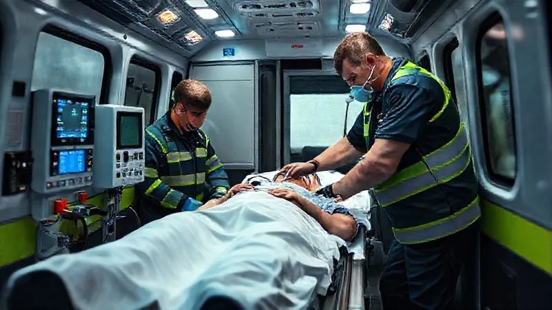
Recognizing the worsening respiratory effort and altered mental status, despite his SpO2 not being critically low by some standards, she immediately called 911. She clearly articulated his baseline, current SpO2 (88% on 2 LPM), the worsening respiratory effort, and his increasing lethargy. When EMS arrived, they found him increasingly somnolent, with an SpO2 now at 85% even after increasing his oxygen to 4 LPM with a simple mask. He was transported to the emergency department.
In the ER, Mr. Henderson was diagnosed with an acute exacerbation of COPD complicated by pneumonia. He required non-invasive ventilation (BiPAP) and aggressive antibiotic therapy. Thanks to his daughter's prompt and astute observation, and her decision to escalate care when his clinical picture deteriorated beyond just the SpO2 number, he avoided intubation and made a full recovery. This case underscores that "low" is relative, and trends, combined with clinical signs, dictate the urgency of intervention. His daughter understood when to escalate care for low SpO2 despite supplemental oxygen.
Communication is Key: What to Tell Healthcare Professionals
When you need to escalate care, effective communication with medical professionals is paramount. Clear, concise, and comprehensive information allows them to quickly understand the situation and make appropriate decisions. I always advise using a structured approach to ensure no critical details are missed.
Here’s what you should be prepared to communicate:
- Patient's Baseline: What is the patient's usual SpO2 range and their typical oxygen requirement?
- Current SpO2 & Oxygen Delivery: What is the current SpO2 reading and what is the exact oxygen flow rate and device (e.g., 90% on 3 LPM nasal cannula)?
- Duration of Low SpO2: How long has the SpO2 been low? Is it a new drop or a persistent issue?
- Associated Symptoms: Describe *all* the other signs you've observed – respiratory effort (faster, harder breathing, accessory muscle use), mental status changes (confusion, lethargy, agitation), skin changes (cyanosis, pallor), and any other symptoms (chest pain, fever, cough).
- Interventions Tried: What have you already done? (e.g., "I checked the tubing, ensured the mask was sealed, and increased oxygen from 2 LPM to 3 LPM, but there was no improvement.")
- Relevant Medical History: Briefly mention key diagnoses (e.g., COPD, heart failure, asthma) that are pertinent to their respiratory status.
Many healthcare systems use the SBAR (Situation, Background, Assessment, Recommendation) framework for clinical communication, which is excellent for organizing your thoughts. For instance, your "Situation" would be the low SpO2 despite oxygen, your "Background" would be their medical history and baseline, your "Assessment" would be the clinical signs you've observed, and your "Recommendation" would be to request an urgent medical review or emergency services. Learning more about SBAR can significantly enhance your ability to communicate effectively in critical situations, as outlined by organizations like the Institute for Healthcare Improvement.
Differentiating Acute vs. Chronic Hypoxemia
Understanding the distinction between acute and chronic hypoxemia is fundamental when determining the urgency of escalation. While both involve low oxygen levels, their implications and management strategies differ significantly.
Chronic Hypoxemia: This refers to persistently low blood oxygen levels over an extended period. It is commonly seen in patients with severe, long-standing lung diseases like COPD or interstitial lung disease. These individuals often adapt to lower SpO2 levels (e.g., 88-92%) and may not show overt signs of distress that someone with acute hypoxemia would. Their bodies have made physiological adjustments, such as increasing red blood cell production, to compensate. For these patients, the goal of oxygen therapy is often to maintain their SpO2 within their acceptable baseline range, typically not aiming for 98-100%, which can sometimes be detrimental.
Acute Hypoxemia: This is a sudden, rapid drop in oxygen levels, often from a normal baseline. It can be caused by acute conditions such as pneumonia, pulmonary embolism, acute respiratory distress syndrome (ARDS), or a severe asthma attack. Acute hypoxemia is far more dangerous because the body has not had time to adapt. Even a SpO2 of 90-92% in someone who normally runs 98% can be a sign of significant acute illness. The focus here is on immediate identification of the cause and rapid intervention to restore oxygenation.
The key takeaway is that when you observe a low SpO2, you must consider the patient's baseline. A SpO2 of 88% might be acceptable for a stable COPD patient on home oxygen, but it's a critical emergency for someone who usually has 98%. It is the acute deviation from the individual's normal, or the worsening of an already compromised state, that mandates urgent escalation, especially when supplemental oxygen isn't helping.
| Feature | Acute Hypoxemia | Chronic Hypoxemia |
|---|---|---|
| Onset | Sudden, rapid | Gradual, long-term |
| Typical SpO2 Target | >94% (generally) | 88-92% (for many conditions) |
| Urgency of Escalation | High, immediate action often needed | Monitored, but less immediate unless acute worsening |
| Associated Symptoms | Often severe distress, rapid mental status changes | May have adapted, fewer acute distress signs unless exacerbated |
The Role of Advanced Monitoring and Diagnostics
While pulse oximetry is an invaluable screening tool, it's just the beginning. When SpO2 remains low despite supplemental oxygen, advanced monitoring and diagnostic tests become crucial for pinpointing the exact cause and guiding definitive treatment. As an industry specialist, I've seen how these tools provide the deeper insights necessary for complex cases.
Beyond the simple pulse oximeter, healthcare providers will often utilize:
- Arterial Blood Gas (ABG): This invasive test provides a precise measurement of oxygen (PaO2), carbon dioxide (PaCO2), and pH in the arterial blood. It offers a much more accurate picture of a patient's oxygenation, ventilation, and acid-base balance than pulse oximetry alone. A low PaO2 confirms hypoxemia, while an elevated PaCO2 indicates respiratory failure (the inability to remove carbon dioxide). Understanding ABG results is a cornerstone of critical care, as detailed in resources like the National Library of Medicine's comprehensive guide on ABG interpretation.
- Chest X-ray: A quick and easy imaging study that can reveal underlying lung pathology such as pneumonia, pulmonary edema (fluid in the lungs), pneumothorax (collapsed lung), or atelectasis (collapsed lung segments).
- Computed Tomography (CT) Scan of the Chest: Provides a more detailed, cross-sectional view of the lungs and surrounding structures, often necessary to diagnose pulmonary embolism, interstitial lung disease, or complex infections.
- Electrocardiogram (ECG/EKG): To assess for cardiac involvement, as heart conditions can mimic or exacerbate respiratory distress. An ECG can detect arrhythmias or signs of myocardial ischemia (heart attack).
- Blood Tests: A complete blood count (CBC) can check for anemia or infection. Inflammatory markers (e.g., C-reactive protein) can indicate systemic inflammation. D-dimer can be used to screen for pulmonary embolism.
These advanced diagnostic tools are typically employed in a hospital setting once a patient has been escalated. They move beyond simply knowing *that* SpO2 is low to understanding *why* it's low, enabling targeted and effective treatment.
Frequently Asked Questions (FAQ)
Question: What if the pulse oximeter reading seems inaccurate? Detailed answer: Pulse oximeters can be inaccurate due to several factors including poor circulation to the finger (cold hands, shock), nail polish (especially dark colors), artificial nails, motion artifact, or carbon monoxide poisoning (which can give falsely high readings). If you suspect inaccuracy, try warming the hand, removing nail polish, using a different finger, or a different device if available. Always correlate the reading with the patient's clinical signs (respiratory effort, mental status). If the patient looks significantly worse than the SpO2 suggests, trust your clinical judgment and escalate.
Question: Can low SpO2 be caused by something other than lung issues? Detailed answer: Yes, absolutely. While most commonly associated with lung problems, severe anemia (not enough red blood cells to carry oxygen), heart failure (poor circulation to deliver oxygenated blood), and certain circulatory conditions can also lead to low SpO2. Additionally, conditions affecting the brain's respiratory drive, such as opioid overdose or stroke, can depress breathing and cause hypoxemia. It's why a comprehensive assessment is always necessary.
Question: Is there a specific SpO2 number that always requires emergency care? Detailed answer: While there isn't a universal 'magic number' because patient baselines vary, an SpO2 consistently below 88% (and certainly below 85%) on supplemental oxygen, particularly if accompanied by worsening clinical signs like severe shortness of breath, confusion, or cyanosis, is a critical emergency for almost any patient. For individuals with no chronic lung disease, an SpO2 below 90% is generally considered a medical emergency. Always consider the trend and the patient's overall condition over an isolated reading.
Question: How can I prepare for a potential SpO2 emergency at home? Detailed answer: If you or a loved one is on home oxygen, preparation is key. Keep a fully charged phone nearby, have emergency numbers (e.g., 911, doctor's office) easily accessible. Ensure your oxygen equipment is regularly maintained and you have backup oxygen supply. Know the patient's baseline SpO2 and their typical oxygen flow. Learn to recognize the critical signs of worsening respiratory distress beyond just the SpO2 number. Discuss an emergency plan with your healthcare provider.
Question: What's the difference between SpO2 and PaO2? Detailed answer: SpO2 (peripheral oxygen saturation) is the percentage of hemoglobin saturated with oxygen, measured non-invasively by a pulse oximeter. It's a good estimate but indirect. PaO2 (partial pressure of arterial oxygen) is the actual pressure of oxygen dissolved in arterial blood, measured invasively via an Arterial Blood Gas (ABG) test. PaO2 is a more direct and accurate measure of oxygenation, providing additional information about ventilation and acid-base balance that SpO2 cannot. Generally, a PaO2 of 80-100 mmHg correlates with an SpO2 of 95-100%, and a PaO2 of 60 mmHg correlates with an SpO2 of about 90%.
Key Takeaways and Final Thoughts
Navigating the complexities of low SpO2, especially when it persists despite supplemental oxygen, demands a blend of vigilance, knowledge, and decisive action. As an experienced industry specialist, I cannot overstate the importance of moving beyond just the numbers and embracing a holistic assessment of the patient's clinical picture. Your ability to recognize subtle changes and act promptly is a powerful tool in ensuring patient safety and positive outcomes.
- SpO2 is a vital sign, not the whole story. Always correlate it with respiratory effort, mental status, and other physical signs.
- Trends matter more than isolated readings. A declining or persistently low SpO2 is a red flag.
- Utilize a structured approach like the ABCDE Checklist to guide your assessment and initial interventions.
- Recognize "red flag" scenarios that demand immediate emergency medical activation.
- Communicate clearly and concisely with healthcare professionals, providing a comprehensive picture.
- Understand the difference between acute and chronic hypoxemia to gauge the urgency of the situation.
Trust your instincts. If a patient "doesn't look right," even if their SpO2 isn't critically low, it's always better to err on the side of caution and seek medical advice. Continuous learning and a proactive approach are your greatest assets in respiratory health. Remember, timely escalation is not a sign of panic, but a hallmark of responsible, expert care. By understanding when to escalate care for low SpO2 despite supplemental oxygen, you empower yourself to make life-saving decisions.
Recommended Reading
- Pro Dancers' Knee Health: How Yoga Prevents Chronic Strain Effectively
- 7 Immune Foods: Optimize Post-Surgery Recovery for Patients
- Elite Cardio: 7 Pillars to Dodge Overtraining Syndrome in Athletes?
- 7 Steps to Build Trust & Cohesion in New Addiction Recovery Groups
- Why Aren't Employees Engaging? 7 Proven Ways to Revitalize Your Wellness Program







0 Comentários: