 By
By 
How to Manage Post-LASIK Epithelial Ingrowth Complications Effectively: An Expert's Perspective
After two decades immersed in the intricate world of ophthalmic surgery, particularly refractive procedures like LASIK, I've witnessed firsthand the profound impact these surgeries have on people's lives. While LASIK consistently delivers remarkable vision correction for millions, it's also true that even with the most meticulous planning and execution, complications can arise. One such challenge, which often causes significant anxiety for both patient and surgeon, is epithelial ingrowth.
Epithelial ingrowth, essentially the migration and proliferation of surface cells under the LASIK flap, can range from an innocuous, self-limiting phenomenon to a sight-threatening condition. It blurs vision, causes glare, and in severe cases, can even melt the corneal tissue, demanding urgent and precise intervention. For patients who have just invested in their vision, discovering this complication can be disheartening and frightening.
My aim with this definitive guide is to illuminate the path forward, providing clarity and actionable strategies on how to manage post-LASIK epithelial ingrowth complications effectively. We’ll delve into the nuances of diagnosis, explore conservative and advanced treatment modalities, discuss crucial post-operative care, and ultimately equip you with the expert insights needed to navigate this challenge with confidence and achieve the best possible visual outcomes.
Understanding Epithelial Ingrowth: The "Why" Behind the Challenge
To truly manage post-LASIK epithelial ingrowth complications effectively, we must first understand its fundamental nature. Epithelial ingrowth refers to the growth of corneal surface cells (epithelium) beneath the LASIK flap. Normally, the epithelial layer forms a protective barrier on the outermost surface of the cornea. During LASIK, a corneal flap is created and lifted, exposing the underlying stromal tissue, which is then reshaped by the laser. After the laser treatment, the flap is repositioned.
The challenge arises when epithelial cells from the corneal surface or the flap's edge migrate and proliferate into the interface between the flap and the underlying stroma. This migration is often triggered by imperfections in flap adherence, residual epithelial cells left inadvertently during surgery, or trauma to the eye post-operatively. These cells, not meant to be in this space, can form nests or sheets, disrupting the smooth interface and scattering light.
Risk factors for developing epithelial ingrowth include re-treatments, thin or irregular flaps, epithelial defects during surgery, and certain systemic conditions. According to a study published in the Journal of Refractive Surgery, the incidence of clinically significant ingrowth requiring intervention is relatively low, typically less than 1-2%, but its potential impact on vision warrants serious attention. Recognizing these underlying causes is the first step in both prevention and effective management.
Early Detection: The Cornerstone of Effective Management
In my experience, the most critical factor in successfully managing epithelial ingrowth is early and accurate detection. Many patients might initially dismiss subtle symptoms, attributing them to normal post-LASIK healing. However, even minor visual disturbances warrant immediate investigation.
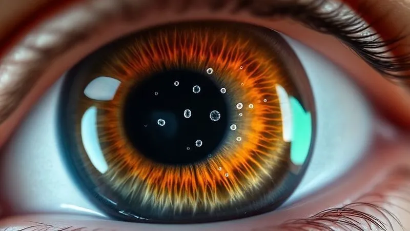
During routine post-LASIK follow-up appointments, I meticulously examine the corneal flap interface using a slit-lamp microscope. Early ingrowth often appears as a translucent, grayish-white haze or sheet-like proliferation, typically starting at the flap edge and slowly progressing inward. It can sometimes have a 'Swiss cheese' appearance with clear lacunae within the cellular sheet.
Patients should be educated to look for persistent or worsening symptoms such as decreased best-corrected visual acuity, glare, halos, or a feeling of something in the eye that doesn't resolve. Any new or worsening visual symptom after the initial healing phase should prompt a visit to your ophthalmologist. Optical Coherence Tomography (OCT) is an invaluable diagnostic tool, offering high-resolution cross-sectional imaging that can precisely map the extent and depth of epithelial ingrowth, guiding treatment decisions with remarkable accuracy.
Conservative Approaches: When to Watch and When to Act
Not all epithelial ingrowth requires immediate surgical intervention. A crucial aspect of how to manage post-LASIK epithelial ingrowth complications effectively is knowing when to observe and when to intervene. Small, non-progressive areas of ingrowth, especially those not affecting the visual axis or causing symptoms, can often be safely monitored.
I typically adopt a 'watchful waiting' approach for Grade 1 or 2 ingrowth (less than 2mm from the flap edge, non-progressive, asymptomatic). Regular follow-up examinations every few weeks are essential to ensure the ingrowth does not progress. In some cases, low-dose topical steroids might be prescribed, though their efficacy in resolving established ingrowth is debated among specialists. The primary goal of conservative management is to avoid unnecessary surgical risks while ensuring patient safety and vision.
However, if the ingrowth shows signs of progression, affects the visual axis, causes significant symptoms (like decreased vision, glare, or foreign body sensation), or leads to flap melt, then intervention becomes necessary. Delaying treatment in such scenarios can lead to more complex and challenging situations. The decision matrix is complex, weighing the risks of surgery against the potential for visual compromise.
| Criteria | Observation (Grade 1-2) | Intervention (Grade 3+) |
|---|---|---|
| Location | Peripheral, >2mm from visual axis | Central, within 2mm of visual axis |
| Symptoms | Asymptomatic or mild, non-progressive glare | Significant vision loss, persistent glare/halos, foreign body sensation |
| Progression | Stable in size and density over months | Growing in size or density, increasing visual impact |
| Corneal Integrity | No signs of flap melt or stromal thinning | Evidence of flap melt, stromal thinning, or necrosis |
Surgical Intervention: Precision and Technique
When conservative measures are insufficient, surgical intervention is the gold standard for how to manage post-LASIK epithelial ingrowth complications effectively. The primary goal is to meticulously remove all epithelial cells from the interface and ensure robust flap adhesion to prevent recurrence. This is a delicate procedure requiring precision and an experienced hand.
The standard surgical approach involves carefully lifting the LASIK flap, similar to the original procedure. Once the flap is re-lifted, the epithelial cells growing on the stromal bed and the undersurface of the flap are gently and thoroughly debrided. I prefer using a combination of blunt and sharp instruments, ensuring complete removal without causing excessive trauma to the underlying stroma. Some surgeons also use a cotton-tipped applicator soaked in 20% ethanol or balanced salt solution to chemically debride the interface, followed by copious irrigation.
After debridement, many surgeons apply Mitomycin-C (MMC) diluted to a specific concentration for a short duration (typically 30-60 seconds) to the stromal bed to inhibit future epithelial cell growth. This must be done with extreme care to avoid corneal toxicity. Following irrigation, the flap is carefully repositioned, and vigorous stroking is performed to ensure tight adhesion. In cases of persistent ingrowth or high risk of recurrence, I may opt for flap suturing to enhance adhesion and prevent further cell migration.
"Effective surgical management of epithelial ingrowth isn't just about removing cells; it's about restoring the corneal architecture and creating an environment where future ingrowth is profoundly discouraged. Precision, patience, and meticulous post-operative care are paramount."
Key Surgical Steps for Epithelial Ingrowth Debridement:
- Flap Re-lift: Carefully re-create the original LASIK flap hinge.
- Interface Irrigation: Thoroughly irrigate the interface with balanced salt solution to loosen epithelial cells.
- Mechanical Debridement: Gently scrape the stromal bed and the undersurface of the flap using a blunt spatula or fine brush to remove all visible ingrowth.
- Chemical Adjuvant (Optional): Apply diluted Mitomycin-C or ethanol to the stromal bed for a controlled duration, followed by extensive rinsing.
- Flap Repositioning & Adherence: Carefully reposition the flap, ensuring proper alignment, and use a wet sponge to firmly stroke the flap, promoting tight adhesion.
- Suturing (Optional): If recurrence risk is high, place fine sutures (e.g., 10-0 nylon) to secure the flap for several weeks.
Advanced Techniques for Recalcitrant Cases
Even with meticulous surgical debridement, epithelial ingrowth can, on rare occasions, recur, presenting a significant challenge. For these recalcitrant cases, I often turn to more advanced techniques. One such method is Phototherapeutic Keratectomy (PTK). After the flap is lifted and debridement is performed, a low-fluence excimer laser can be applied to the stromal bed to ablate any remaining microscopic epithelial cells and create a smoother, less hospitable surface for ingrowth. This can be particularly effective in preventing recurrence, especially when combined with MMC.
Another strategy for persistent ingrowth involves a repeat flap lift and debridement, sometimes with a longer application of MMC or a different flap fixation technique. In extremely rare and severe cases where significant flap melt or stromal scarring has occurred, a corneal transplant (penetrating keratoplasty or deep anterior lamellar keratoplasty) might be considered, though this is truly a last resort. The key is to be adaptable and persistent in the treatment strategy, customizing it to the specific characteristics of the ingrowth and the patient's corneal health.
Case Study: Sarah's Recurrent Ingrowth
Sarah, a 38-year-old teacher, initially underwent LASIK three years prior. She presented with a recurrence of Grade 3 epithelial ingrowth affecting her central vision, despite a previous debridement procedure six months earlier. The ingrowth was dense and causing significant glare, impacting her ability to read and drive at night. After a thorough diagnostic evaluation, I opted for a repeat flap lift and debridement, but this time, I incorporated a 45-second application of 0.02% Mitomycin-C to the stromal bed, followed by extensive irrigation. Additionally, I placed two interrupted 10-0 nylon sutures to secure the flap for six weeks, providing enhanced stability. Post-operatively, Sarah received an intensive regimen of topical steroids and antibiotics. At her three-month follow-up, there was no evidence of recurrence, and her vision had improved significantly to 20/20, with a dramatic reduction in glare. This case underscored the importance of aggressive, multi-modal treatment for persistent ingrowth.
Post-Operative Care and Prevention of Recurrence
The success of any intervention for epithelial ingrowth doesn't end with the surgery; it extends deeply into meticulous post-operative care and diligent prevention of recurrence. This phase is as critical as the surgical procedure itself. Immediately after debridement, patients are typically placed on an intensive regimen of topical corticosteroids to minimize inflammation and topical antibiotics to prevent infection. The steroid regimen is carefully tapered over several weeks or even months, depending on the severity of the ingrowth and the response to treatment.
I often use a bandage contact lens (BCL) for the first week post-op to provide comfort and promote epithelial healing, which helps to seal the flap edge and reduce the risk of epithelial cell migration. Patient education is paramount: they must understand the importance of avoiding eye rubbing, adhering strictly to the medication schedule, and attending all follow-up appointments. Any trauma to the eye can potentially re-open the flap interface and trigger a recurrence.
Long-term monitoring is also essential. Even after successful treatment, I schedule regular check-ups for several months, sometimes up to a year, to ensure there are no subtle signs of new cell growth. Vigilance from both the patient and the ophthalmologist is the best defense against recurrence. Understanding corneal health and potential complications is key for long-term management.
Navigating Patient Expectations and Emotional Support
Beyond the clinical aspects, effectively managing post-LASIK epithelial ingrowth complications also involves significant patient communication and emotional support. A patient who develops a complication after elective surgery can feel anxious, frustrated, and even regretful. As their physician, it's my responsibility to address these concerns with empathy and transparency.
I always ensure patients fully understand the nature of epithelial ingrowth, the proposed treatment plan, and the realistic expectations for recovery and visual outcome. It's crucial to acknowledge their feelings and provide reassurance that while this is a setback, it is a manageable one with high success rates when treated appropriately. Setting realistic expectations is vital; while vision often improves significantly, it may not always return to the pre-complication level, especially in severe or recurrent cases. Providing clear, consistent information helps to build trust and reduce anxiety. The psychological impact of surgical complications should never be underestimated, and a supportive approach is part of holistic care.
The Future of Ingrowth Management: Innovations on the Horizon
The field of ophthalmology is continuously evolving, and the quest for better ways to manage post-LASIK epithelial ingrowth complications effectively is ongoing. Researchers are exploring various avenues, from novel pharmacological agents to advanced imaging techniques. One promising area is the development of targeted anti-proliferative drugs that could be applied topically to prevent epithelial cell migration without affecting overall corneal health. These could potentially reduce the need for surgical intervention in mild to moderate cases.
Newer generations of femtosecond lasers are also being investigated for their ability to create more precise and stable flap architectures, potentially reducing the initial risk of ingrowth. Furthermore, advanced diagnostic tools with even higher resolution and specificity might allow for the detection of ingrowth at its earliest, most microscopic stages, enabling intervention before it becomes clinically significant. As a community, we are always pushing the boundaries to make refractive surgery even safer and more predictable. Research into novel therapeutic strategies for corneal complications is advancing rapidly.
| Aspect | Current Approaches | Future Innovations |
|---|---|---|
| Diagnosis | Slit-lamp, OCT | AI-enhanced OCT, molecular biomarkers for early detection |
| Prevention | Meticulous surgical technique, proper flap adherence | Targeted anti-migratory topical agents, bio-engineered flap interface coatings |
| Treatment | Flap lift & debridement, MMC/ethanol, sutures, PTK | Novel selective epithelial ablation technologies, gene therapy for cell proliferation control |
Frequently Asked Questions (FAQ)
Q: What are the long-term visual impacts of untreated epithelial ingrowth? Untreated epithelial ingrowth can lead to significant and permanent visual impairment. It can cause progressive blurring, ghosting, glare, and halos as the cells disrupt the corneal optics. In severe cases, the proliferating epithelial cells can release enzymes that digest and melt the corneal tissue (flap melt), leading to irregular astigmatism, corneal scarring, and even perforation, potentially requiring a corneal transplant. Early intervention is crucial to prevent these severe long-term consequences.
Q: Can epithelial ingrowth resolve on its own? Very small, peripheral, and non-progressive areas of epithelial ingrowth (often referred to as Grade 1) can sometimes remain stable or even regress spontaneously without intervention. However, once ingrowth begins to progress, especially if it's near the visual axis or causing symptoms, it is highly unlikely to resolve on its own and typically requires active management. Monitoring is key to differentiate between stable and progressive cases.
Q: Is epithelial ingrowth more common with certain LASIK techniques? Historically, microkeratome-created flaps might have had a slightly higher incidence of ingrowth due to less precise flap creation compared to femtosecond laser-created flaps. However, modern microkeratomes are highly refined. The primary factors influencing ingrowth are less about the tool itself and more about surgical technique, the presence of epithelial defects during surgery, and flap adhesion post-operatively, regardless of the initial flap creation method. Re-treatment procedures are generally associated with a higher risk of ingrowth.
Q: What if I have ingrowth in both eyes? While less common, bilateral epithelial ingrowth can occur. The management strategy for each eye would typically be determined independently, based on the severity, location, and progression of the ingrowth in that specific eye. If surgical intervention is required for both, it would usually be performed sequentially to allow for healing and monitoring of the first eye before proceeding with the second, minimizing bilateral vision impairment during recovery.
Q: How soon after LASIK can ingrowth appear? Epithelial ingrowth can appear anywhere from days to several months after LASIK. Most cases become clinically apparent within the first few weeks to three months post-operatively. However, late-onset ingrowth can also occur, sometimes triggered by minor trauma or flap manipulation years after the initial surgery. This is why consistent follow-up and patient awareness of symptoms are important for an extended period after LASIK.
Key Takeaways and Final Thoughts
Managing post-LASIK epithelial ingrowth complications effectively requires a blend of clinical expertise, meticulous diagnostic skills, and a patient-centered approach. From early detection to precise surgical intervention and diligent post-operative care, each step plays a crucial role in safeguarding visual outcomes and ensuring patient satisfaction.
- Vigilance is Key: Early detection through regular follow-ups and patient awareness of symptoms is paramount.
- Tailored Treatment: Not all ingrowth requires surgery; conservative management is appropriate for stable, asymptomatic cases.
- Surgical Precision: When needed, meticulous debridement, potentially with adjunctive therapies like MMC, is crucial for success.
- Prevent Recurrence: Intensive post-operative care and long-term monitoring are essential to prevent the return of ingrowth.
- Empathy Matters: Supporting patients emotionally and managing their expectations is integral to comprehensive care.
As an industry specialist, I can assure you that while epithelial ingrowth can be a daunting complication, advancements in diagnosis and treatment mean that excellent outcomes are achievable. Remain proactive, communicate openly with your eye care professional, and trust in the process. Your vision is precious, and with the right approach, we can overcome these challenges together. For further reading and to find a qualified ophthalmologist, I recommend consulting resources from the American Academy of Ophthalmology.
Recommended Reading
- Unlock Vitality: Safe Physical Activity for Beginners Over 50
- Long-Distance Hiker's Foot Pain: 7 Proven Strategies for Lasting Relief
- High-Stakes Stress & Illness: Your 5-Step Immunity Shield
- 7 Proven Strategies for Addressing Patient Refusal of Cancer Screenings
- Chronic Inflammation & Immunity: 7 Steps to Reclaim Control







0 Comentários: