 By
By 
How to Differentiate Between IBS and Subtle IBD in Complex Cases?
For over two decades in the challenging landscape of digestive health, I've witnessed countless individuals grappling with chronic gut symptoms. Many arrive at my door with an ‘IBS’ label, yet their journey feels incomplete, their symptoms persistent, and their unease palpable. This feeling, this persistent doubt, often whispers a truth: sometimes, what presents as a straightforward functional disorder might, in fact, be something far more insidious and organic – a subtle form of Inflammatory Bowel Disease (IBD).
The problem is profound: distinguishing between Irritable Bowel Syndrome (IBS) and subtle IBD in complex cases is one of the most demanding diagnostic puzzles we face in gastroenterology. Patients endure prolonged suffering, frustration, and a diminished quality of life when the underlying pathology remains undiagnosed or misdiagnosed. The nuances are often missed, leading to inappropriate treatments and a delay in addressing potentially progressive inflammatory conditions.
In this comprehensive guide, I aim to equip you with the expert frameworks, diagnostic insights, and clinical wisdom necessary to confidently approach these challenging scenarios. We will explore the critical red flags, advanced diagnostic tools, and the art of clinical judgment required to accurately determine how to differentiate between IBS and subtle IBD in complex cases, ensuring patients receive the precise care they desperately need.
The Elusive Overlap: Why Differentiation is So Challenging
The primary reason why differentiating IBS from subtle IBD is so difficult lies in the significant overlap of their presenting symptoms. Both conditions can manifest with abdominal pain, bloating, altered bowel habits (diarrhea, constipation, or mixed), and fatigue. This symptomatic mimicry creates a diagnostic minefield, often leading to an initial IBS diagnosis when, in reality, early-stage IBD is simmering beneath the surface.
Understanding IBS: A Functional Disorder
IBS is fundamentally a disorder of gut-brain interaction, characterized by chronic abdominal pain related to defecation, or associated with a change in stool frequency or form. It's defined by the Rome IV criteria, focusing on symptom patterns rather than structural abnormalities. The pathophysiology involves visceral hypersensitivity, altered gut motility, dysbiosis, and disturbances in the gut-brain axis. Crucially, IBS is not associated with inflammation or structural damage to the bowel wall. This is a key distinction, yet one that can be deceptively absent in early IBD.
Understanding Subtle IBD: The Early, Tricky Stages
Subtle IBD refers to the early or atypical presentations of Crohn's disease or ulcerative colitis. These are the cases where the inflammation is microscopic, patchy, or confined to areas not easily accessed by standard colonoscopy, such as the small bowel. Patients might not present with the classic ‘red flag’ symptoms like significant weight loss, bloody stools, or severe anemia initially. Microscopic colitis, for example, shares many symptoms with IBS-D but involves distinct inflammatory changes visible only on biopsy. Early Crohn's disease can manifest with vague abdominal discomfort and fatigue long before overt strictures or fistulas develop. These are the cases that truly test our diagnostic acumen.
Key Insight: "The absence of overt inflammation on routine blood tests or initial imaging does not always mean the absence of underlying organic disease. Subtle signs demand deeper investigation."
Clinical Red Flags: Beyond the Standard IBS Checklist
While IBS and IBD share symptoms, certain clinical features should always raise a red flag, prompting a more aggressive pursuit of an IBD diagnosis. In my experience, overlooking these subtle cues is where the most significant diagnostic errors occur.
- Unexplained Weight Loss or Anemia: While patients with IBS might lose weight due to dietary restrictions, significant, unintentional weight loss that cannot be attributed to diet changes is a powerful indicator of malabsorption or systemic inflammation. Similarly, iron deficiency anemia, especially if persistent and not explained by menstrual blood loss, should always trigger an IBD workup.
- Nocturnal Symptoms: Diarrhea or abdominal pain that consistently wakes a patient from sleep is highly atypical for IBS. IBS symptoms typically resolve or significantly lessen during sleep. Nocturnal symptoms suggest an underlying inflammatory or organic process that isn't influenced by conscious control or daily stressors.
- Family History of IBD: Genetics play a significant role in IBD. If a first-degree relative (parent, sibling, child) has Crohn's disease or ulcerative colitis, the patient's risk is substantially elevated. This factor alone should lower the threshold for advanced investigations.
- Abnormal Inflammatory Markers (Even if Subtle): While overtly elevated C-reactive protein (CRP) or erythrocyte sedimentation rate (ESR) are clear IBD indicators, even slightly elevated or fluctuating levels in a patient with chronic symptoms should not be dismissed as insignificant. Similarly, fecal calprotectin, a marker we will discuss in detail, is invaluable here.
- Persistent or Progressive Symptoms: IBS symptoms tend to fluctuate in intensity and pattern. If symptoms are relentlessly progressive, worsening over time, or failing to respond to conventional IBS therapies, it's a strong signal to reconsider the diagnosis. This includes new onset of symptoms in older adults (over 50), which is less typical for IBS.
These red flags are not definitive diagnostic criteria on their own, but they are crucial pieces of the puzzle that, when present, should significantly increase suspicion for IBD, guiding the next steps in the diagnostic process.
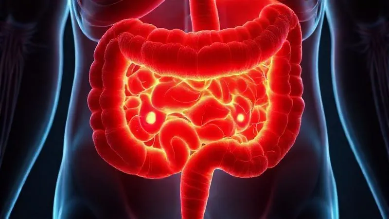
Advanced Diagnostic Tools: Peering Beyond the Surface
When clinical suspicion is high, or red flags are present, relying solely on symptom-based diagnosis is insufficient. We must employ advanced diagnostic tools that can detect the subtle inflammatory changes characteristic of early IBD.
Calprotectin and Lactoferrin: The Non-Invasive Sentinels
Fecal calprotectin and lactoferrin are proteins released by neutrophils into the gut lumen during inflammation. They are incredibly useful non-invasive biomarkers for differentiating inflammatory bowel disease from functional gastrointestinal disorders like IBS. Elevated levels strongly suggest intestinal inflammation, making them excellent screening tools. While a very high level (e.g., >250 µg/g for calprotectin) is highly indicative of active IBD, even slightly elevated levels (e.g., 50-200 µg/g) in a symptomatic patient should not be ignored. A normal fecal calprotectin (<50 µg/g) makes IBD highly unlikely, though not entirely impossible in very select cases.
Key Insight: "Fecal calprotectin, even when slightly elevated, demands attention in a complex case. It's a powerful non-invasive pointer towards the need for endoscopic investigation." According to a review in the World Journal of Gastroenterology, fecal calprotectin is a highly sensitive and specific marker for intestinal inflammation.
Endoscopy with Biopsies: The Gold Standard, Reimagined
Colonoscopy with ileoscopy remains the gold standard for diagnosing IBD. However, in complex cases, it's not just about performing the procedure; it's about performing it with an IBD-suspecting mindset. This means:
- Thorough Inspection: Scrutinizing the entire colon and terminal ileum for subtle erythema, loss of vascular pattern, aphthous ulcers, or granular mucosa that might be missed on a cursory examination.
- Multiple Biopsies: Taking numerous biopsies from all segments of the colon (even if appearing normal), including the terminal ileum. Microscopic colitis, for instance, is diagnosed solely on biopsies showing specific inflammatory patterns (e.g., collagenous or lymphocytic colitis) in macroscopically normal mucosa. Early Crohn's can also present with patchy microscopic inflammation.
- Experienced Pathologist: Ensuring the biopsies are reviewed by a pathologist experienced in gastrointestinal pathology, who can identify the subtle architectural changes and inflammatory infiltrates that distinguish early IBD from normal tissue.
Case Study: Maria's Persistent Abdominal Pain
Maria, a 34-year-old marketing executive, had been diagnosed with IBS-D five years prior. She experienced intermittent abdominal pain, bloating, and urgent diarrhea, often exacerbated by stress. Despite trying various diets and medications, her symptoms persisted, and she occasionally reported mild fatigue. Her initial blood tests and a routine colonoscopy two years ago were normal. However, a recent fecal calprotectin test, performed due to a subtle, unexplained weight loss of 5 pounds, came back at 110 µg/g – elevated but not dramatically high. This prompted a repeat colonoscopy with extensive biopsies. This time, the gastroenterologist specifically targeted areas of the terminal ileum and ascending colon that, while appearing mostly normal, had a slightly granular texture. The pathology report revealed microscopic aphthous ulcerations and focal chronic inflammation consistent with early Crohn's disease. Maria's seemingly 'IBS' symptoms were, in fact, the subtle whispers of an emerging IBD, finally caught through diligent investigation.
Advanced Imaging: MRI Enterography and Capsule Endoscopy
When symptoms strongly suggest small bowel involvement, but standard endoscopy is negative, advanced imaging techniques become crucial. Small bowel Crohn's disease, in particular, can be notoriously difficult to diagnose early.
- MRI Enterography (MRE): This non-invasive imaging technique provides detailed views of the small bowel, looking for inflammation, strictures, fistulas, and wall thickening. It's excellent for visualizing the entire small intestine and surrounding mesentery, which is often affected in Crohn's.
- Capsule Endoscopy: A tiny camera swallowed by the patient, which captures images as it passes through the entire small bowel. It's highly sensitive for detecting mucosal lesions like erosions or ulcers that might be missed by other modalities. However, it cannot take biopsies and may miss subtle transmural inflammation.
The Role of Biomarkers and Genetics: Unlocking Deeper Clues
Beyond inflammation markers, other biological and genetic indicators can provide further insights, particularly in challenging diagnostic scenarios.
Serological Markers: ASCA and pANCA
Perinuclear anti-neutrophil cytoplasmic antibodies (pANCA) and anti-Saccharomyces cerevisiae antibodies (ASCA) are serological markers often used in conjunction with clinical findings. ASCA is more commonly associated with Crohn's disease, while pANCA is more often found in ulcerative colitis. While not diagnostic on their own, their presence can increase the probability of IBD, especially when used in panels. They are not perfect, having limitations in sensitivity and specificity, but in a complex case where other markers are equivocal, they can offer additional supportive evidence.
Genetic Predisposition: When Family History Echoes
While IBD is not a purely genetic disease, genetic factors play a significant role in susceptibility. Specific genes, such as NOD2/CARD15, have been strongly linked to Crohn's disease. While routine genetic testing isn't a primary diagnostic tool for IBD, understanding a patient's genetic predisposition, especially in the context of a strong family history, can inform the level of vigilance required. Research published in Nature Reviews Gastroenterology & Hepatology highlights the complex genetic architecture of IBD, involving hundreds of susceptibility loci.
| Feature | IBS (Typical) | Subtle IBD (Early) | IBD (Active) |
|---|---|---|---|
| Calprotectin | <50 µg/g | 50-250 µg/g (often elevated) | >250 µg/g |
| CRP | Normal | Normal to slightly elevated | Elevated |
| Endoscopy | Normal mucosa | Microscopic inflammation, subtle changes | Gross inflammation, ulcers |
| Weight Loss | Rare/minimal | Possible, unexplained | Common |
The Art of Clinical Judgement: Beyond the Labs and Scans
While diagnostic tools are indispensable, the seasoned clinician's judgment remains paramount. Synthesizing all available information – patient history, physical examination, laboratory results, and imaging findings – into a cohesive diagnostic picture is an art. This is particularly true when you are trying to understand how to differentiate between IBS and subtle IBD in complex cases, where no single test provides a definitive answer.
Longitudinal Observation and Symptom Progression
Sometimes, the diagnosis isn't immediately obvious. In such situations, a period of watchful waiting combined with careful longitudinal observation is necessary. Monitoring symptom progression, response to empirical treatments, and repeating non-invasive markers like fecal calprotectin after a few months can provide crucial clarity. A patient initially diagnosed with IBS might, over time, develop more overt IBD symptoms or consistently elevated inflammatory markers, necessitating re-evaluation.
Response to Treatment: A Diagnostic Clue
The patient's response to treatment can also serve as a diagnostic clue. If a patient with an IBS diagnosis fails to respond to conventional IBS therapies (e.g., fiber, antispasmodics, low FODMAP diet, neuromodulators), it should prompt a re-evaluation for underlying organic disease. Conversely, if a patient with suspected IBD responds dramatically to anti-inflammatory medications, it strengthens the IBD diagnosis. This iterative process of diagnosis and therapeutic trial is often part of managing complex gut issues.
Navigating Patient Communication and Expectations
Diagnosing chronic conditions, especially when there's uncertainty, requires sensitive and clear communication with patients. The emotional toll of living with chronic digestive symptoms is immense, and a prolonged diagnostic journey can exacerbate anxiety and distrust.
Empowering Patients with Knowledge
I always advocate for empowering patients with knowledge. Explain the diagnostic process, the reasons for further testing, and the potential for a nuanced diagnosis. Discussing the overlap between IBS and IBD openly helps manage expectations and fosters a partnership in their healthcare journey. Providing resources from reputable organizations like the Crohn's & Colitis Foundation can be invaluable.
The Multi-Disciplinary Approach
Complex digestive cases often benefit from a multi-disciplinary approach. Collaborating with dietitians, psychologists (especially for gut-directed hypnotherapy or CBT for IBS management), and other specialists ensures a holistic view of the patient's condition. This integrated care can address not only the physical symptoms but also the significant psychological impact of chronic illness, regardless of the final diagnosis.
When to Re-evaluate: The Dynamic Nature of Gut Health
It's crucial to understand that a diagnosis, especially in the realm of functional versus organic gut disorders, is not always static. The gastrointestinal tract is a dynamic system, and conditions can evolve. What might appear as classic IBS today could, months or years down the line, reveal itself to be early IBD.
Key Insight: "A negative colonoscopy today does not guarantee the absence of IBD tomorrow if symptoms persist or worsen. Vigilance and re-evaluation are cornerstones of responsible gastroenterological care." This proactive approach is supported by guidelines from organizations like the American College of Gastroenterology, which emphasize ongoing assessment.
Therefore, if a patient with an established IBS diagnosis experiences a significant change in symptoms – new red flags emerge, symptoms become more severe, or they fail to respond to previously effective treatments – a full re-evaluation is warranted. This might involve repeating inflammatory markers, imaging, or even endoscopy. Maintaining an open mind and a willingness to revisit a diagnosis are critical skills for any expert in digestive health, especially when tackling the intricate question of how to differentiate between IBS and subtle IBD in complex cases.
Frequently Asked Questions (FAQ)
Question 1: Can IBS turn into IBD? No, IBS cannot 'turn into' IBD. They are distinct conditions with different underlying pathologies. IBS is a functional disorder without inflammation, while IBD is a chronic inflammatory disease. However, it's common for early or subtle IBD to be initially misdiagnosed as IBS due to symptom overlap. The appearance of IBD after an IBS diagnosis usually indicates that the IBD was always present but initially undetected.
Question 2: How often should I be re-evaluated if I have an IBS diagnosis but symptoms are worsening? If your IBS symptoms are worsening, or if new symptoms (especially red flags like unexplained weight loss, nocturnal symptoms, or blood in stool) develop, you should consult your gastroenterologist promptly. There's no fixed timeline, but any significant, persistent change warrants a re-evaluation, which might include repeat blood tests, stool tests (like fecal calprotectin), or even imaging/endoscopy.
Question 3: Are there any specific dietary interventions that help differentiate? While dietary interventions like a low FODMAP diet can significantly improve IBS symptoms, they typically do not resolve the inflammation associated with IBD. If severe symptoms persist despite strict adherence to an appropriate diet for IBS, it could be a clue that an underlying inflammatory condition is present. However, diet alone is not a diagnostic tool for differentiation; it's more about symptom management.
Question 4: What's the role of stress in confusing IBS and IBD symptoms? Stress can significantly exacerbate symptoms in both IBS and IBD due to the gut-brain axis. In IBS, stress can trigger flares of pain and altered bowel habits. In IBD, stress can worsen symptoms and potentially contribute to flares of inflammation. This overlap makes differentiation challenging, as both conditions can appear to worsen with stress. However, stress alone does not cause the inflammation seen in IBD. Addressing stress is important for both conditions but doesn't rule out IBD.
Question 5: If my fecal calprotectin is slightly elevated but endoscopy is normal, what's next? A slightly elevated fecal calprotectin (e.g., 50-200 µg/g) with a normal endoscopy presents a true diagnostic dilemma. This scenario often requires careful clinical correlation. Options include repeat fecal calprotectin in 3-6 months, further imaging of the small bowel (like MRI enterography) if small bowel Crohn's is suspected, or consideration of microscopic colitis which requires specific targeted biopsies. Sometimes, a period of watchful waiting with close symptom monitoring is appropriate, especially if symptoms are mild. A study in Gastroenterology highlights the complexity of interpreting such results.
Key Takeaways and Final Thoughts
- Embrace Vigilance: Never settle for an IBS diagnosis if red flags persist or symptoms progress. Always maintain a high index of suspicion for subtle IBD.
- Leverage Non-Invasive Markers: Fecal calprotectin is your primary screening tool. Don't dismiss even slightly elevated levels in complex cases.
- Optimize Endoscopic Technique: When endoscopy is performed, ensure thorough inspection and multiple, targeted biopsies, especially from the terminal ileum and any suspicious areas.
- Integrate Advanced Imaging: For suspected small bowel involvement, MRI enterography and capsule endoscopy are invaluable.
- Trust Clinical Judgement: Synthesize all data, observe longitudinally, and consider treatment response as diagnostic clues.
- Communicate Empathetically: Guide patients through the diagnostic journey with clarity and compassion.
The journey to accurately differentiate between IBS and subtle IBD in complex cases is often intricate and demanding. It requires a blend of diagnostic acumen, an understanding of advanced tools, and above all, a commitment to the patient's well-being. By adopting these expert strategies, we move beyond mere symptom management to precise diagnosis, paving the way for effective, targeted treatment and a significantly improved quality of life for our patients. Remember, in the realm of gut health, vigilance is not just a virtue; it's a necessity.
Recommended Reading
- Mastering Focus: 7 Strategies for Joint Pain in Demanding Roles
- 5 Strategies: Overcoming Unexpected Plateaus in Chronic Pain Rehabilitation
- Fact vs. Fiction: 7 Ways to Counter Douching Misinformation in Young Women
- The Secret to Enduring Protection: How Booster Shots Enhance Long-Term Immunity
- 5 Proven Strategies: Solving Low-Income Clients' Healthy Eating Adherence?







0 Comentários: